CELL PHYSIOLOGY
- This is the study of the functions of cell structures.
Membrane Structure and Properties
- A membrane is a surface structure which encloses the cell and organelles. Membranes regulate the flow of materials into out of the cell or organelle.
- Examples of membranes: cell membrane, tonoplast (membrane surrounding the vacuole), nuclear membrane, mitochondrial membrane, chloroplast membrane etc.
The Cell Membrane
- It has three layers, two protein layers and a phos-pholipid layer sandwiched in between the two.
Diagram
Properties of Cell Membrane
- Semi-permeability. – It has small pores allowing for the passage of molecules of small size into and out of the cell. Cell Wall however allows all materials to pass through it hence it is referred to as being Permeable.
- Sensitivity to Changes in Temperature and pH – Extreme temperature and pH affects the cell membrane since it has some protein layers. Such changes alter the structure of the membrane affecting its normal functioning.
- Possession of Electric Charges – it has both the negative and positive charges helping the cell to detect changes in the environment. These charges also affect the manner in which substances move in and out of the cell
Physiological Processes
- The ability of the cell to control the movement of substances in and out of the cell is achieved through physiological processes such as Diffusion, Osmosis and Active Transport.
Diffusion
- This is a process by which particles move from a region of high concentration to a region of low concentration.
Practical Activity 1
To demonstrate diffusion using potassium permanganate (VII)
- The difference in concentration of particles between the region of high concentration and the region of low concentration is known as the diffusion gradient.
Role of Diffusion in Living Organisms
- Absorption of Materials
- Mineral salts in the soil enter the root by diffusion since their concentration in the soil is greater than in the root hair cells.
- Digested food (glucose and amino acids) diffuse across the wall of the ileum into the blood for transport to rest of the body.
- Gaseous Exchange in Plants and Animals
- In both plants and animals, respiratory gases (oxygen and Carbon (IV) oxide) are exchanged through simple diffusion depending on their concentration gradient.
- Excretion of Nitrogenous Wastes
- Transport of Manufactured Food form Leaves to other Plant Parts.
Factors Affecting Diffusion
- Diffusion Gradient
- A greater diffusion gradient between two points increases the rate of diffusion.
- Surface Area to Volume Ratio
- The higher the ratio the greater the rate of diffusion and the lower the ratio the lower the rate.
- This means that small organisms expose a large surface area to the surrounding compared to large organisms.
- Small organisms therefore depend on diffusion as a means of transport of foods, respiratory gases and waste products.
Diagrams
- Thickness of Membranes and Tissues
- The thicker the membrane the lower the rate of diffusion because the distance covered by the diffusing molecules is greater. The thinner the membrane, the faster the rate.
- Size of the Molecules
- Small and light molecules diffuse faster than large and heavy molecules.
- Temperature
- Increase in temperature increases the energy content in molecules causing them to move faster.
Osmosis
- This is the process where solvent molecules (water) move from a lowly concentrated solution (dilute) to a highly concentrated solution across a semi-permeable membrane.
Diagram fig 4.6
- The highly concentrated solution is known as Hypertonic Solution.
- The lowly concentrated solution is called Hypotonic solution.
- Solution of the same concentration are said to be Isotonic.
- Osmosis is a special type of diffusion because it involves the movement of solvent (water) molecules from their region of high concentration to region of low concentration across a semi permeable membrane.
Practical activity 2
Practical activity 3
Osmotic Pressure
- This is the pressure which needs to be applied to a solution to prevent the inward flow of water across a semi permeable membrane. This is the pressure needed to nullify osmosis.
- Osmotic pressure is measured using the osmometer.
Osmotic Potential
- This is the measure of the pressure a solution would develop to withdraw water molecules from pure water when separated by a semi permeable membrane.
Water Relations in Animals
- Cell membrane of the animal cell is semi permeable just like the dialysis/visking tubing.
- Cytoplasm contains dissolved sugars and salts in solution form.
- If an animal cell e.g. a red blood cell is placed in distilled water (hypotonic solution), water flows in by osmosis.
- The cell would swell up and eventually burst because the cell membrane is weak. The bursting of the red blood cell when placed in hypotonic solution is called Haemolysis.
- If a similar red blood cell is placed in a hypertonic solution, water is drawn out of the cell by osmosis. The cell will shrink by a process called Crenation.
- Body fluids surrounding the cells must therefore have same concentration as to that which is found inside the cell.
Diagrams
Water Relations in Plants
- When a plant cell is placed in a hypotonic solution it gains water by osmosis and distends outwards.
- As the cell gains more water, its vacuole enlarges and exerts an outward pressure called turgor pressure. As more water is drawn in, the cell becomes firm and rigid and is said to be turgid.
- The cell wall in plant cell is rigid and prevents the cell from bursting unlike the case in animal cells.
- The cell wall develops a resistant pressure that pushes towards the inside. This pressure is equal and opposite the turgor pressure and is called wall pressure.
Diagrams
- When a plant cell is placed in hypertonic solution, water molecules move out of the cell into the solution by osmosis. The cell shrinks and becomes flaccid.
- If the cell continues to lose more water, plasma membrane pulls away from the cell wall towards the center.
- The process through which plant cells lose water, shrink and become flaccid is called plasmolysis.
- Plasmolysis can be reversed by placing a flaccid cell in distilled water and this process is called deplasmolysis.
Study Question 5
Practical Activity 4
Wilting
- When plants lose water through evaporation and transpiration, cells lose turgidity, shrink and the plant droops. This is called wilting.
- If water supply from the soil is inadequate, plants do not recover hence permanent wilting.
Study Question 6
Role of Osmosis in Organisms
- Absorption of water from the soil
- Root hair cells of plants absorb water from the soil by osmosis.
- Support
- Cells of herbaceous plants, which are less woody, absorb water, become turgid hence support.
- Opening and closing of the stomata
- During the day, guard cells synthesize glucose, draw in water, become turgid hence open the stomata.
- During the night, they lose turgidity since there is no photosynthesis. As a result, they shrink thus closing the stomata.
- Feeding in insectivorous plants
- These plants are able to change their turgor pressure on the leaves which close trapping insects which are digested to provide the plant with nitrogen.
- Osmoregulation
- In the kidney tubules, water is reabsorbed back to the body by osmosis.
Factors Affecting Osmosis
- Concentration of Solutions and Concentration Gradient. The greater the concentration gradient between two points, the faster the rate of osmosis.
- Optimum Temperature as long as it does not destroy the semi-permeability of the membrane.
Active Transport
- This is the process that moves substances across cell membranes against a concentration gradient.
- This process requires energy to move these substances across cell membranes and involves carriers.
- Substances such as amino acids, sugar and many ions are taken in by living organisms through active transport.
Role of Active Transport
- Re-absorption of sugars and useful substances by the kidney
- Absorption of some mineral salts by plant roots
- Absorption of digested food from the alimentary canal into the blood stream
- Accumulation of substances in the body to offset osmotic imbalance in arid and saline environment
- Excretion of waste products from body cells
Factors Affecting Active Transport.
- Oxygen concentration.
- Change in pH.
- Glucose concentration.
- Temperature.
- Enzyme inhibitors.
NB/ Any factor affecting energy production affect the rate of active transport.
Revision Questions.
Cell Specialization, Tissues, Organs and Organ Systems
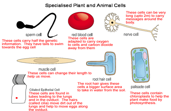
- Cell specialization
- This is where cells are modified to perform specific functions. Such cells are said to be specialized.
- Examples include the sperm cell which has tail for swimming and the root hair cell which is extended creating large surface area for water absorption.
- Tissues.
- These are cells of a particular type that are grouped together to perform the same function.
Animal tissues include;
- Epithelial tissue – which is a thin continuous layer of cells for lining and protection of internal and external surfaces.
- Skeletal – it is a bundle of elongated cells with fibres that can contract. Its contraction and relaxation brings about movement.
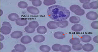
Blood tissue – this is a fluid containing red blood cells, white blood cells and platelets.
It transports many substances and protects the body against infections.
- Connective tissue – made up of strong fibres that connect other tissues and organs holding them together.
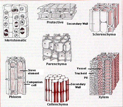
Plant tissues include:
- Epidermal tissue of a plant – this is a single layer of cells protecting the inner tissues of the plant.
- Palisade tissue – this is a group of cells rich in chloroplasts containing chlorophyll. They absorb light energy during photosynthesis.
- Parenchyma tissue – it is made thin walled irregularly shaped cells. They store water and food.
- Vascular bundle – consists of the xylem and phloem. Xylem conducts water and mineral salts while phloem conducts food substances.
- Organs
- Many tissues become specialized and grouped together to perform a functional unit called the organ.
- Examples of organs in plants include; roots, leaves, flowers and stem.
- In animals they include heart, lungs, kidney, brain, stomach and the liver.
- Organ systems.
- This is made of several organs whose functions are coordinated and synchronized to realize an effective action is called an organ system. Examples include; digestive, circulatory, excretory, respiratory, reproductive and nervous system.
Revision Questions
MICROSCOPE
Microscope Parts & Function
Parts of the Microscope
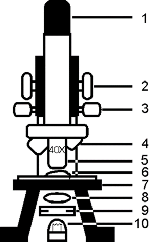
| 1. Eyepiece | Contains a magnifying lens that focuses the image from the objective into your eye. |
| 2. Course Adjust | For focusing under low magnification |
| 3. Fine Adjust | For focusing under high magnification or low |
| 4. Low Power Objective | For large specimens or overview |
| 5. High Power Objective | For detailed viewing or small specimens |
| 6. Specimen on glass slide | What you want to look at |
| 7. Stage | Supports specimen in correct location to lens |
| 8. Condenser | Focuses the light on specimen |
| 9. Diaphragm (iris or disc) | Regulates amount of light and contrast |
| 10. Light Source | Illuminates the specimen for viewing |
Handling and Care of the Microscope
The following rule should be observed:
- Use both hand when carrying the microscope. One hand should hold the base and the other holds the limb.
- Never place the microscope too close to the edge of the bench.
- Do not touch the mirror and the lenses with the fingers.
- Clean dirty lenses using soft tissue.
- Clean other parts using a soft cloth.
- Do not wet any part of the microscope.
- Make sure the low power clicks into position in line with the eye piece before and after use.
- Always store the microscope in a safe place free from dust and moisture.
Using the Microscope
- Place microscope on the bench with the stage facing away from you.
- Turn the low power objective lens until it clicks into position.
- Ensure the diaphragm is fully open.
- Look through the eyepiece with one eye. Adjust the mirror to ensure maximum light can pass through.
- Place the slide containing the specimen on the stage and clip it into position. Make sure the slide is at the centre of the field of view.
- Again look through the eyepiece while adjusting the mirror to ensure maximum light reach the specimen.
- Use the coarse adjustment knob to bring the low power objective lens to the lowest point. While viewing through the eyepiece, turn the coarse adjustment knob gently until the specimen comes into focus.
- Use the fine adjustment knob to bring the image into sharp focus.
- Make a drawing of what you see.
- For higher magnification, turn the medium power into position and adjust the focus using the coarse knob. Use the fine adjustment knob for sharper focus.
- For even large magnifications, turn the high power objective lens into position. In this case use only the fine adjustment knob to bring details into sharper focus.
Magnification
- Magnification of the object viewed under the microscope is calculated by;
Magnification = Eye Piece Lens Magnification X Objective Lens Magnification.
- If the eyepiece lens has the magnification of x5 and the low power objective lens has a magnification of x10, the total magnification is 5×10=50.
Study Question 1
Fill the table below.
| Eye piece lens maginification | Objective lens magnification | Total magnification |
| X5 | X4 | |
| X10 | X5 | |
| X10 | X100 | |
| X40 | X600 | |
| X10 | X100 |
Practical Activity 1
Cell Structures as Seen Under the Light Microscope
- The following cell organelles can be seen under the light microscope.
- Cell wall.
- Cell membrane
- Cytoplasm
- Nucleus
- Vacuole.
- Chloroplasts.
Diagrams- plant and animal cells
The Electron Microscope.
- It is more powerful than the light microscope.
- It can magnify up to 500,000 times and has high resolving power.
- The high resolving power of the electron microscope enables it to separate objects which lie close to one another.
- Electron microscope uses a beam of electrons instead of light to illuminate the object.
Study Question 2
Practical Activity 2
Cell Structures as Seen Under the Electron Microscope
Diagrams – Plant and Animal Cells
The Cell Organelles
- Cell membrane (Plasma Membrane).
- It has three layers i.e. one layer of phospho-lipid layer sandwiched between two protein layers.
- It is flexible with pores and ahs the following main functions.
- Encloses all the cell contents.
- It allows selective movement of substances into and out of the cell since it is semi-permeable.
Diagram
- Cytoplasm
- It is s fluid medium in which chemical reactions take place.
- It has some movement called cytoplasmic streaming.
- It contains organelles, starch, glycogen, fat droplets and other dissolved substances.
- Nucleus
- It has double membrane called the nuclear membrane.
- The membrane has pores allowing passage of materials into and out of the cell.
- Nucleus has a fluid called nucleoplasm in which the nucleolus and chromatin are suspended.
- Nucleolus manufactures ribosomes while chromatin contains the hereditary material.
- Mitochondria(Mitochondrion)
- They are sausage shaped and are the respiratory sites.
- Mitochondrion has two membranes. Inner membrane is greatly folded into cristae to increase the surface area for respiration.
- Cells that require a lot of energy have large number of mitochondria e.g. muscle cell, sperm cell, kidney cell etc.
Diagram
- Endoplasmic Reticulum (ER)
- Some endoplasmic reticulums have granules called Ribosomes on their surfaces hence referred to as rough endoplasmic reticulum.
- Others do not contain ribosomes hence the name smooth endoplasmic reticulum.
- Rough endoplasmic reticulum transport proteins while the smooth endoplasmic reticulum transports lipids.
Diagrams
- Ribosomes
- They are spherical in shape and form the site for protein synthesis.
- Lysosomes
- They contain lytic enzymes which break down large molecules, destroy worn out organelles or even the entire cell.
- Golgi Bodies (Golgi apparatus)
- Their function is to package and transport glyco-proteins.
- They are also associated with secretion of synthesized proteins and carbohydrates.
Diagram
- Centrioles
- They are rod shaped structures that are used in cell division and in the formation of cilia and flagella.
- Plant cells lack the Centrioles.
- Chloroplasts
- They are egg shaped and contain two membranes.
- Chloroplast has chlorophyll which traps light energy to be used during photosynthesis.
- Vacuoles
- This are sacs filled with a fluid called cell sap.
- Animal cells contain small vacuoles while plant cells have large vacuoles.
- Sap vacuoles store sugars and salts.
- Food vacuole store and digest food while contractile vacuoles excrete unwanted materials from the cell.
- Cell wall
- It is a rigid outer cover of the plant cells made of cellulose.
- It gives the plant cell a definite shape while providing mechanical support and protection.
- Cell wall also allows water, gases and other materials to pass through it.
Study Question 3
Differences between Plant and Animal Cells
Preparation of Temporary Slides
Practical Activity 3
Estimation of Cell Sizes.