REPRODUCTION IN PLANTS AND ANIMALS
- This is the process by which mature individuals produce offsprings.
- There are two types of reproduction.
- Sexual reproduction which involves male and female gametes
Diagram
- Asexual reproduction where no gametes are involved.
Diagram
Importance of Reproduction
- Procreation
This ensures that a species does not become extinct.
- Quality improvement
Reproduction allows for mixing of genetic materials bringing
about variations.
These variations are important tools in the refinement of quality of offsprings.
Cell Division
- Life in all living things start as a single cell as a spore or as a zygote.
- The cells have to divide further to give rise to make cells.
- Cell division starts with division of the nucleus (chromosome) and then the cytoplasm.
Chromosomes
- These are microscopic thread like structure within cells that carries the molecule deoxyribonucleic acid (DNA)—the hereditary material that influences the development and characteristics of each organism.
- Each chromosome is made up of two parallel strands called chromatids.
- Chromatids are joined together at one point by the centromere.
Diagram
- Each cell has a fixed number of chromosomes e.g. each human body cell has 46 chromosomes.
- Chromosomes occur in pairs in the nucleus. A member of each pair is called homologous chromosomes.
- Homologous chromosomes are similar in appearance, size, and shape but their genetic constitution may be different.
- Genes are found along the length of the chromosomes.
- Genes are very tiny and made up of a chemical substance called DNA (De oxy Ribonucleic Acid)
- DNA determines the characteristics of the offspring.
- There are two types of cell division
- Mitosis
- Meiosis
Mitosis
- In this type of cell division, each cell divides into two daughter cells each having the same number of chromosomes as the parent cell.
- Mitosis occurs in series of stages i.e.
- Interphase
- Prophase
- Metaphase.
- Anaphase
- Telophase.
1)Interphase
During this stage the following activities take place within the cell in preparation of the division.
- Synthesis of new cell organelles such as ribosome’s, centrioles, mitochondria and Golgi apparatus.
- Multiplication of genetic material so that each daughter cell will have same number of chromosomes as the parent cell.
- Build up of enough energy stores in form of ATP (Adenosine Triphosphate) during respiration. This energy is important to see the cell through the process of division.
- At this stage the chromosomes are not clearly visible.
Diagrams
2)Prophase
The following events take place in this stage.
- Centrioles separate and move to opposite poles of the cells.
- Spindle fibres begin to form
- Nuclear membrane begins to break down and nucleolus disappears.
- Chromosomes thicken and shorten and they can be stained easily hence become visible.
Diagram
3)Metaphase
- Nuclear membrane disappears and chromosomes are free in the cytoplasm.
- Spindle fibres lengthen and attach to the centrioles at both poles.
- Chromosomes align themselves at the equator and are attached to the spindle fibres by their centromere.
Diagram
- Anaphase
- Chromatids separate at the centromere and migrate to opposite poles. This is brought about by the shortening of the spindle fibres.
- Spindle fibres begin to disappear.
- In animal cells, cell membrane begins to constrict towards the end of anaphase.
Diagram
- Telophase
- Chromatids collect together at the two opposite poles of the spindle.
- Nuclear membrane forms around each set of chromatids and are now referred to as chromosomes.
- Cytoplasm divides into two hence the formation of two daughter cells.
- Chromosomes become less distinct.
In animal cells, division of cytoplasm is by constriction of cell membrane.
In plant cells, a cell plate forms within the cytoplasm and grows to separate the cell into two.
Diagrams
Significance of Mitosis
- Forms basis for asexual reproduction e.g. budding and spore formation.
- Causes cell growth when the cells formed increase in number and size.
- Ensures genetic constitution of the offspring is the same as the parents.
- Replaces damaged and dead cells in the body.
Meiosis
- This involves two divisions of the parental cell resulting into four daughter cells.
- First meiotic cell division involves the separation of the homologous chromosomes. It is referred to as Reduction division because the numbers of chromosomes are reduced by half.
- In the second stage, the sister chromatids are separated and it is referred to as Equatorial division
- Each daughter cell has half the number of chromosomes (haploid n) as the parent cell.
- This takes place in the reproductive organs of animals (testis and ovary) and plants (anthers and ovary).
- Meiosis is divided into same series of stages as in mitosis.
- The phases are given names as in mitosis but each is followed by I or II.
First Meiotic Division
Interphase I
The cell prepares for division by the following.
- Replication of chromosomes.
- Synthesis of new cell organelles.
- Build up of energy.
Prophase I
- Nucleolus disappears.
- Centrioles move to opposite poles.
- Chromosomes shorten and thicken becoming more visible.
- Homologous chromosomes lie side by side in the process of synapsis forming pairs called bivalents.
- Homologous chromosomes may become coiled around each other with their chromatids remaining in contact at points called chiasmata.
NB/. During chiasma formation homologous chromosomes may exchange genetic material during crossing over. These genetic exchanges are important because they bring about variations in offsprings.
Metaphase.I
- Nuclear membrane disappears.
- Homologous chromosomes as a bivalent move to the equator of the cell.
- Spindle fibres are fully formed and get attached to the chromosomes at the centromere.
- Homologous chromosomes orientate towards different poles.
Diagram
Anaphase I
- Homologous chromosomes separate and migrate to the opposite poles with their centromeres leading. This is brought about by the shortening of the spindle fibres.
Diagram
Telophase I
- Cell divides across the middle when the chromosomes reach the poles.
- At the end of meiosis I homologous chromosomes are separated.
Diagram
Second Meiotic Division.
In this stage the sister chromatids are separated from each other.
Interphase II
- Cells go into a short interphase.
Prophase II
- Chromosomes become shorter and thicker.
- New spindle fibres are formed.
Metaphase.II
- Chromosomes align at the equator of the cell.
- Spindle fibres attach to their centromeres.
- Chromosomes orientate themselves towards the opposite poles.
Anaphase II
- Sister chromatids separate from each other.
- Spindle fibres shorten pulling them to the opposite poles.
Telophase II
- Spindle fibres disappears
- Nucleolus reappears and nuclear membrane forms around each set of chromatids.
- Chromatids uncoil and become threadlike.
- Cytoplasm divides.
- Four cells are formed (tetrad).
- Each cell has haploid (n) number of chromosomes.
Significance of Meiosis
- Gamete formation (sperms and ova) forming basis for sexual reproduction
- Provides opportunities for genetic variations during crossing over
Similarities between mitosis and meiosis
- Both take place in plants and animals.
- Both involve division (multiplication) of cells.
Differences between meiosis and mitosis
| Meiosis | Mitosis. |
| Homologous chromosomes associate with each other. | No association of homologous chromosomes |
| Takes place in 2 nuclear divisions. | Takes place in one nuclear division. |
| 4 daughter cells are produced each haploid (n) | 2 daughter cells are produced each diploid (2n) |
| Occurs in reproductive organs leading to gamete formation. | Occurs in somatic (body) cells leading to growth. |
| Chiasma formation takes place leading to crossing over hence variation | No chiasma formation therefore no crossing over hence no variation. |
Asexual Reproduction
- This is the production of offsprings from a single organism without fusion of gametes.
- This type of reproduction involves mitosis.
Types of Asexual Reproduction
- Binary fission in amoeba, plasmodium and bacteria
- Sporulation in rhizopus
- Budding in yeasts
- Binary fission in amoeba
- When there is enough food and favourable temperature and pH, a mature amoeba divides into two.
- During binary division, in amoeba, internal reorganization of molecules necessary for structural construction takes place.
- Nucleus first divides mitotically (Karyogamy) into two followed by the division of the cytoplasm (Cytogamy)
Diagrams
- Sporulation in Rhizopus
- This is the formation of spores in substrates like the bread to form bread moulds
- A spore is a microscopic reproductive unit which contains a nucleus and a small amount of cytoplasm.
- Spores are produced by bacteria, most fungi, mosses and ferns.
- Rhizopus has a vegetative body called the mycelium.
- Mycelium is made up of many branched threads called hyphae.
- Horizontal hyphae are called stolons.
- Vertically growing ones are called sporangiophore.
- Tips of sporangiophore swell up to form the sporangia (sporangium).
- Sporangia are the spore bearing structures. When fully mature, sporangium wall burst releasing the spores. If spores land on a suitable medium, they germinate and develop into other rhizopus.
- Rhizopus uses structures called rhizoids for anchorage and to obtain nutrients from the substrate.
Diagrams
Budding in Yeast
Under favourable conditions such as plenty of sugar, moisture, oxygen and optimum temperature, the yeast cell reproduces asexually by budding.
- A projection of bud forms on the parent cell.
- Nucleus divides into two.
- One nuclei moves into the new bud.
- Bud grows in size and forms new cell organelles. Later the bud separates off.
Diagrams
Sexual Reproduction in Plants
- In flowering plants the flower is the reproductive organ.
Structure and Function of a Flower
- A flower is made up of a flower stalk (pedicel) and a receptacle.
- Attached to the receptacle are four groups of floral structures i.e.
- Calyx (sepals)
- Corolla/petals
- Androecium – male parts
- Gynoecium – female parts
- Calyx (sepals)
- Made up of the sepals which are usually green.
- If sepals are fused they form gamosepalous calyx.
- If they are free, they form polysepalous calyx.
- Calyx protects the inner parts of the flower especially during bud development.
- Some flowers have sepal like structures below the calyx called the epicalyx.
- Corolla/petals
- It’s made up of the petals which are brightly coloured, large and conspicuous especially in insect pollinated flowers.
- If fused – gamopetalous.
- If free – polypetalous
- Androecium – male parts
- Made up of one or more stamens
- Satmen is made up of the filament and anthers.
- Another has four pollen sacs containing pollen grains.
- Pollen grains contain the male gametes.
Diagrams
- Gynoecium – female parts
- It may contain one or more carpels
- A carpel consists of the ovary, the style and the stigma.
- Ovary contains the ovules.
- Ovaries are described as epigynous, hypogynous or perigynous depending on the place they occur in the flower.
- Epigynous (inferior) ovary
- Ovary is located within the receptacle.
- All other floral parts occur above it such as in the apple flowers.
Diagram
- Hypogynous (superior) ovary
- Ovary is above the receptacle and other floral parts such as in hibiscus.
Diagram
- Perigynous ovary
- The receptacle surrounds the carpel.
- All other floral parts arise around the ovary such as in roses.
Diagram
The gynoecia can also be grouped into different types dependi.ng on the number of carpels present i.e. monocarpous or syncarpous.
Monocarpous Gynoecium
- It has only one carpel e.g. in beans.
Diagram
Polycarpous Gynoecium
- It has two or more carpels. It is divided into two.
- Apocarpous gynoecium
- The carpels are free e.g in roses and bryophyllum.
Diagrams
- Syncarpous gynoecium
- The carpels are fused together such as in hibiscus.
Diagrams
Terms Used in Describing a Flower
- Complete flower – has all the four floral parts; calyx, corolla, androecium and gynoecium.
- Incomplete flower – has one or two floral parts missing.
- Unisexual flower – a flower with only one of the reproductive parts either male or female flower.
- Staminate flower – male flower.
- Pistillate flower – female flower.
- Monoecious plant – bears both male and female parts of the flower.
- Dioecious plants – the plant is either male or female e.g. in paw paw.
- Hermaphrodite or bisexual flower – has both the male and female parts.
- Regular or actinomorphic flower – a flower that can be divided into tow similar halves by any vertical section passing through the center i.e. radial symmetry such as in morning glory.
- Irregular or zygomorphic flower – can be divided into two similar halves on one particular plane only i.e. bilateral symmetry e.g. in clotalaria.
- Pedicillate flower- flower with a stalk.
- Solitary flower – are flowers occurring singly.
- Inflorescence – flowers that grow in clusters.
- Essential parts of the flower – are the androecium and gynoecium.
- Non essential floral parts – are the calyx and corolla.
Pollination
This is the transfer of pollen grains from the anther to the stigma.
Types of Pollination
- Self pollination. – Transfer of pollen grains from the anther to the stigma of the same flower.
- Cross Pollination – transfer of pollen grains from the anther of one flower to the stigma of another flower but of the same species.
Agents of Pollination
- Insect
- Wind
Adaptations of Insect Pollinated Flowers (Entomophilous)
- Flowers are large, conspicuous with brightly coloured petals and inflorescence to attract insects.
- Flowers are scented and produce nectar to attract insects.
- Pollen grains are relatively large, heavy, rough or sticky so as to stick on to the body of the sticks.
- They have small and firmly attached anthers to a firm filament.
- Stigmas are small, sticky and contained within the flower. This ensures that pollen grains from the body of an insect stick onto it.
- Flowers have a tubular or funnel shaped corolla, landing platforms and honey guides.
Adaptations of Wind Pollinated Flowers (Anemophilous)
e.g. maize and other grasses
- Small flowers with inconspicuous petals, bracts or inflorescence.
- Flower structure is simple and flowers have no particular shape.
- Stigmas are long, feathery and hang outside the flower to trap pollen grains.
- Pollen grains are small, smooth and light to be easily carried by the wind.
- Flowers are not scented and lack nectar.
- Anthers are large and loosely attached to a flexible filament to be easily released when the wind blows.
Diagram of a grass flower
| Filament |
Features and Mechanisms Hindering Self Pollination and Self Fertilization
- Heterostyly – condition whre the stigma na d style have different arrangements e.g. coconut flowers have shorter stamens than pistils hence pollen grains from the anthers cannot reach the stigma.
Diagram
- Self sterility or incompatibility – condition where pollen grains of a flower fail to germinate if they land on the stigma of the same flower.
- Protogyny and Protandry – condition where either male parts of a flower mature before the female ones.
Protandry – stamen mature before the stigma e.g.in sunflower.
Protogyny – stigma matures before the anthers mature e.g. in maize.
- Dioecious plants and presence of features that promote cross pollination such as brightly coloured petals which attract insects hence cross pollination.
Fertilization in Flowering Plants
Fertilization in plants is the fusion of the male and female nuclei in the embryo sac.
- Male gamete is contained in the pollen grain produced in the anther.
Diagram
Female gamete (egg cell) is found in ovules contained in the embryo sac.
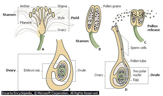
Process of Fertilization
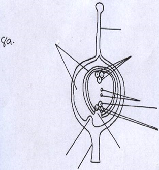
- Pollen grains land stick to the stigma and germinates to form pollen tube, which grows through the tissue of the style towards the ovary
- The generative nucleus undergoes mitosis, forming 2 male nuclei
- The pollen tube gets into the embryo sac through the micropyle; pollen tube nucleus disintegrates, creating a passage for the male nuclei.
- The egg cell fuses with one of the two male nuclei to form a diploid zygote. The zygote undergoes mitosis to form an embryo
- The two polar nuclei fuse with the second male nucleus to form a triploid nucleus.
- The triploid nucleus forms the endosperm. The two con current fertilization incidents are collectively referred to as double fertilization
Seed and Fruit Development
- Some changes occur to the ovary, ovule and the entire flower after fertilization.
- Calyx dries and falls off or may persist.
- Petals and stamens wither and fall off.
Development of the Seed
- Zygote undergoes mitotic division to become the embryo (plumule and radicle) and one or two cotyledons.
- Primary endosperm nucleus develops into the endosperm.
- Ovule forms the seeds.
- Ovary develops into a fruit.
- Integuments become the seed coat (testa).
- Testa has got a scar (hilum) which is the attachment point to the placenta.
- A seed a tiny opening called the micropyle which allows water into the seed during germination.
- Water is withdrawn from the seed from about 80% to 15% by mass making the seed dry and hard.
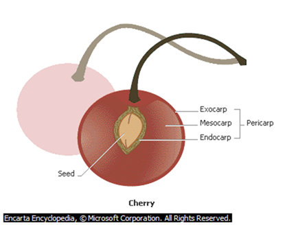
Development of Fruits
- A fruit is a fully grown fertilized ovary containing fully developed seeds.
- This is brought about by the hormones gibberellins and occurs after fertilization.
- As the ovules develop into seeds, the rest of the ovary develops into the fruit wall or the pericarp.
- Pericarp has two scars indicating the points of attachment to the style and to the receptacle.
- Pericarp has three layers; epicarp/exocarp (outer most), mesocarp (middle) and the endocarp (innermost).
- In some fruits such as pineapples and bananas fruit formation takes place without fertilization. This is called parthenocarpy.
- False fruits are formed when other parts of the flower such as the receptacle enlarge and enclose the ovary e.g. in pineapples, apple, straw berry and cashew nut.
Classification of Fruits
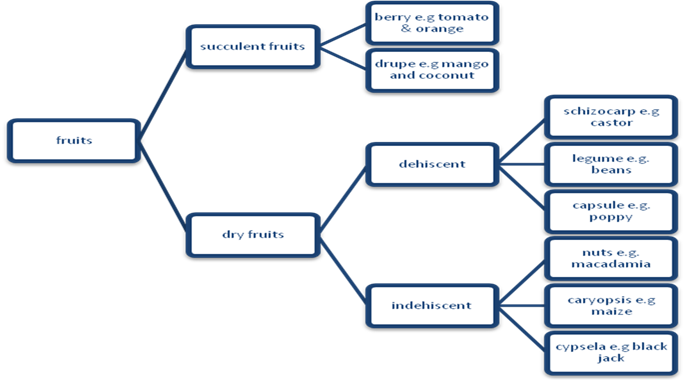
Succulent fruits
They are divided into berry and drupe.
Berry – has a succulent pericarp divided into epicarp, mesocarp and endocarp e.g. orange, tomato, passion fruit, melon, paw paw etc.
Diagram
Drupe – they have a thin epicarp, fleshy or fibrous mesocarp and a very hard endocarp enclosing the seeds. In mango the fleshy edible part is the mesocarp while in coconut the mesocarp is a fibrous cover just before the hard endocarp.
Diagram
Dry Fruits
- They are divided into dehiscent and indehiscent.
Dry Dehiscent fruit
They dehisce to release their seeds. They are divided into;
- Legume e.g beans
Diagram
- Capsule e.g poppy
Diagram
- Schizocarp e.g. castor.
Diagram
Dry indehiscent fruits
- These do not dehisce.
- They include;
- Caryopsis – pericarp and seed coat are fused together to form a thin covering round the seed e.g. maize.
Diagram
- Cypsela – it’s a one seeded e.g. the blackjack.
Diagram
- Nut – the pericarp becomes hard and woody and it is separate from the seed coat e.g. macadamia.
Diagram
Placentation
- This is the arrangement of the ovules in an ovary. They include;
- Marginal Placentation.
- Ovules are attached to the placenta in a row e.g. peas in a pod.
Diagram
- Basal placentation
- Placenta is formed at the base of the ovary. Ovules are attached to it sunflower and sweet pepper.
Diagram
- Axile Placentation
- The edges of the carpels fuse together to form a central placenta in the axile.
- Ovules are arranged on the placenta.
- The ovary is divided into a number of loculi by the walls of the carpel e.g. in orange
Diagram
- Parietal Placentation
- Edges of the carpels fuse together and dividing walls disappear leaving a loculus.
- Placentas from each carpel appear as a ridge on the ovary wall and have numerous ovules on them e.g. in paw paw.
Diagram
- Free central placentation
- Edges of carpels fuse together and the dividing walls disappear leaving one loculus.
- Placenta appears at the center and have numerous ovules on it e.g. in primrose
Diagram
Adaptations of Fruits to Various Agents of Dispersal
- Water dispersal
- Such seeds and fruits enclose air in them to lower their density for buoyancy;
- They are fibrous/ spongy to lower the density for buoyancy;
- Have impermeable seed coat or epicarp to prevent water from entering during flotation so as to avoid rotting;
- The seeds can remain viable while in water and only germinate while on a suitable medium;
- Wind dispersal
- They are light; and small; to be easily carried by wind currents due to lower density;
- Have developed extension (Parachute like structures and Wing like structures) which create a larger surface area; so as to be kept afloat in wind currents e.g. sonchus and jacaranda
- In some a Perforated capsule is usually loosely attached to a long stalk which is swayed away by wind scattering seeds;
- Animal dispersal
- Brightly colored to attract animals
- Fleshy to attract animals; e.g. mangoes, passion fruits, oranges, tomatoes etc.
- aromatic /scented to attract animals;
- The seed coats are hard and resistant to digestive enzymes; the seeds are therefore dropped away in feaces/droppings e.g. passion fruit and tomatoes.
- Some have hook like structures to attach on animals fur e.g. blackjack
d) Self dispersal
- They have weak lines (sutures) on the fruit wall (pod), along which they burst open to release seeds, which get scattered away from the parent plant e.g. in legumes such as peas and beans.
SEXUAL REPRODUCTION IN ANIMALS
- This involves gamete fusion.
- The male produces the male gamete (sperms) and the female produces the female gamete (ovum/ova).
- The gametes are produced in special organs called gonads i.e. the testes and ovaries.
- The sperm fuses with the ovum to form a zygote through a process called fertilisation the gametes are haploid and the zygote is diploid.
- Fertilisation may be internal or external.
External Fertilisation in Amphibians
- The female lays eggs and the male sheds sperms on them (to fertilise them). This is only possible in water.
- Many eggs are released to increase the chances of survival since bacteria and other organisms can eat fertilised eggs.
- Eggs are also in long strands of slippery jelly like substance, which offer the eggs protection.
- This substance separates the eggs from each other allowing for good aeration.
- It also attaches the eggs to water plants and makes them buoyant.
Internal Fertilisation
- This occurs in reptiles, birds and mammals where fertilisation occurs within the body of the female.
- Sperms are introduced into the female’s body.
- Few eggs are produced because there are high chances of fertilisation and the gametes/zygote receive further protection.
- In most mammals, some chameleons and some snakes the fertilised eggs develop into young ones within the body of the female. They give birth to young ones.
Study Question 8
Reproduction in Mammals
- Mammals have internal fertilization where eggs are laid or develop within the female’s body in the uterus.
- The egg laying mammals (monotremes) they are said to be oviparous such as the platypus.
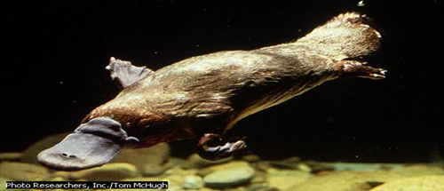
Platypus
The duck-billed platypus, Ornithorhynchus anatinus, found only in eastern Australia, belongs to an unusual group of egg-laying mammals called monotremes. It lives in streams, rivers, and occasionally lakes. The duck-billed platypus feeds on bottom-dwelling aquatic insect larvae, which it finds by probing the streambed with its pliable, sensitive bill.
- In marsupials such as the kangaroo the zygote does not develop fully within the uterus but completes development in the pouch.
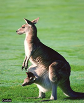
Mother Kangaroo and Baby
Kangaroos are a type of mammal called a marsupial. Baby marsupials are unable to survive on their own when they are born, so they must live in a pouch on their mother’s belly. A newborn kangaroo, called a joey, stays in its mother’s pouch for about six months, where it feeds on her milk.
- The ability to give birth to young ones as in placental mammals is called viviparity.
- Mammals have mammary glands, which produce milk on which the young ones are fed. Parental care is highly developed in mammals.
Reproduction in Human beings
Structure and Function of The male Reproductive System
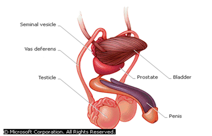
Male Reproductive System
The organs of the male reproductive system enable a man to have sexual intercourse and to fertilize female sex cells (eggs) with sperm. The gonads, called testicles, produce sperm. Sperm pass through a long duct called the vas deferens to the seminal vesicles, a pair of sacs that lies behind the bladder. These sacs produce seminal fluid, which mixes with sperm to produce semen. Semen leaves the seminal vesicles and travels through the prostate gland, which produces additional secretions that are added to semen. During male orgasm the penis ejaculates semen.
- Testes are found outside the abdominal cavity in the scrotal sac. This position provides a cooler environment for sperm production since sperms develop best at lower temperature than that of the body.
- Testis is made up of highly coiled tubes called seminiferous tubules whose inner lining has actively dividing cells which give rise to sperms.
- Between the seminiferous tubules are interstitial cells, which produce the male hormones (androgens).
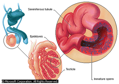
Internal View of Male Reproductive System
The reproductive anatomy of the male human is largely external. Beginning at puberty, sperm are produced within seminiferous tubules of the testicles, a pair of glands that reside in a pouch called the scrotum. The external location of the scrotum keeps the temperature of sperm slightly below body temperature, which is necessary for their healthy development and survival. From each testicle, sperm migrate to a long, coiled tube known as the epididymis, where they are stored for one to three weeks until they mature. Also located outside the body is the penis, the erectile organ responsible for the excretion of urine and the transfer of sperm to the vagina of the female. Just before ejaculation during sexual arousal, mature sperm travel from the epididymis, a coiled tube behind each testicle, through a long duct called the vas deferens. Sperm leave the body in semen, a fluid produced by the seminal vesicles.
- Seminiferous tubules unite to form the epididymis, which is about 6m long and highly coiled. It stores the sperms.
- It’s connected to the sperm duct/vas deferens. Sperm duct connects the epididymis to the urethra, which is the ejaculatory duct.
- Seminal vesicles provide an alkaline fluid, which contains nutrients for the sperms.
- Prostate gland secretes an alkaline substance to neutralise the vaginal fluids. It also activates the sperms.
- Cowper’s glands secrete an alkaline fluid that neutralizes the acidity along the urethra.
- All these fluids combine with the spermatozoa to form the semen.
- Since the urethra serves both passage of urine and semen it is said to be urino-genital in function.
- The penis is erectile and made of spongy tissue, muscle and blood vessels.
- Once erect, the penis is able to penetrate the vagina in order to deposit sperms into the female’s reproductive tract.
Study question 9 and Practical.
Structure and Function of The Female Reproductive System.
Diagram
- The internal sex organs of the female consist of the vagina, uterus, fallopian tubes (or oviducts), and ovaries.
- The vagina is a flexible tube-shaped organ that is the passageway between the uterus and the opening in the vulva. Because during birth the baby travels from the uterus through the vagina, the vagina is also known as the birth canal.
- The woman’s menstrual flow comes out of the uterus and through the vagina.
- When a man and a woman engage in vaginal intercourse, the penis is inserted into the vagina.
- The cervix is located at the bottom of the uterus and includes the opening between the vagina and the uterus. It secretes a plug of mucus, which prevents entry of pathogens into the uterus during pregnancy.
- The uterus is a muscular organ that has an inner lining (endometrium) richly supplied with blood vessels and glands. During pregnancy, the uterus holds and nourishes the developing foetus.
- Although the uterus is normally about the size of a fist, during pregnancy it is capable of stretching to accommodate a fully developed foetus, which is typically about 50 cm (about 20 in) long and weighs about 3.5 kg (about 7.5 lbs).
- The uterine muscles also produce the strong contractions of labour.
- At the top of the uterus are the pair of fallopian tubes (oviduct) that lead to the ovaries.
- The two ovaries produce eggs, or ova (the female sex cells that can become fertilized), and female sex hormones, primarily oestrogen and progesterone.
- The fallopian tubes have finger like projections at the ends near the ovaries that sweep the egg into the fallopian tube after it is released from the ovaries.
- Movement of ovum is also aided by the smooth muscles of the oviduct.
- If sperm are present in the fallopian tube, fertilization (conception) may occur and the fertilized egg will be swept into the uterus by cilia (hair like projections inside the fallopian tube).
Practical
The Human Sperm
- Are formed in the seminiferous tubules of testes by meiosis.
- Final products of meiosis enter the sertoli cells where they are nourished and undergo maturation.
- Mature sperms leave for epididymis where they are stored.
- A mature sperm has an ovoid head, short neck, middle piece and a tail.
Diagram
- Head has a large nucleus carrying the genetic material, which is haploid (n).
- At the tip of the head there is the acrosome containing lytic enzymes. These enzymes digest the wall of ova.
- The short neck contains centrioles.
- Middle piece has a large number of mitochondria, which provide with the energy required for propulsion of the sperm to reach the ova.
- The tail propels the sperm forward by its side-to-side lashing action.
Formation of The Ova
- In females egg formation begins in the ovary of the foetus before birth unlike in males where production of sperms starts at puberty.
- At birth there are about 70,000 potential egg cells in the ovaries of a baby girl.
- A layer of ovary cells called primary follicles, which provide them with nourishment, encloses them.
- Only about 500 of them develop into ova during puberty. During puberty the primary follicles grow to become Graafian follicle.
- At ovulation, the Graafian follicle bursts open to release a mature ovum surrounded by a layer of cells.
Diagram
- A mature ovum is spherical in shape with a diameter of about 0.2 mm.
- It has a large haploid nucleus surrounded by a nuclear membrane.
- Nucleus is within the cytoplasm enclosed by the plasma membrane. Vitelline membrane surrounds the plasma membrane.
Study Question 11
Fertilisation
- Process where the nucleus of a male gamete fuses with the nucleus of a female gamete to form a zygote.
- This takes place in the upper part of he oviduct after copulation. Sperms are drawn up by suction through the cervix into the uterus. They swim up to the oviduct using their tails.
- Very many sperms are released but only one is required to fertilise the ovum.
- The ovum releases chemical substances, which are neutralised by those released by the acrosome.
- When the ovum comes into contact with the egg the acrosome bursts releasing lytic enzymes, which dissolve the egg membranes.
- The acrosome turns inside out forming a filament, which is used to penetrate the eggs.
Diagrams
- The Vitelline membrane undergoes a change, which stops any other sperm from entering the ovum.
- Once inside the cytoplasm the head bursts to release the male nucleus, which then fuses with the female nucleus to form a diploid zygote.
- After ovulation the ovum can remain viable for 8-24 hours before it dies.
- The sperm can remain viable for 2-3 days in the female reproductive tract.
Study Question 12
Implantation
- This is the attachment of the blastocyst to the walls of the uterus by the villi.
- After fertilisation, the zygote undergoes various mitotic divisions as it moves down the oviduct. Its movement is aided by cilia in the oviduct and by the contractions of the smooth muscles lining the oviduct.
- By the time it reaches the uterus it has formed a hollow structure of cells called blastocyst.
- Movement of the zygote from the oviduct to the time it is implanted takes about 7 days.
Diagrams
- Sometime the zygote may fail to move down to the uterus and gets implanted into the walls of the oviduct. This condition is referred to as ectopic pregnancy.
Formation of Placenta
- During implantation the blastocyst differentiates into three layers, chorion, amnion and allantois.
Diagram
- Chorion is the outermost and it has finger like projections called chorionic villi. These villi grow into the endometrium. During the early stages of embryo development, villi form the sites for material exchange between the embryo and maternal blood vessels.
- Amnion surrounds the embryo forming an amniotic cavity. Amniotic cavity contains the amniotic fluid, which suspends the foetus providing it with support. It also acts as a shock absorber hence protecting it against mechanical injury.
- The chorionic villi, allantois and the endometrium form the placenta.
- The embryo is attached to the placenta by a tube called the umbilical cord.
- When the placenta is fully formed, the embryo becomes the foetus at about three months of pregnancy.
The Role of The Placenta
- This is a temporary organ found only in placental mammals. It is the only organ in animals composed of cells derived from two different organisms; the foetus and the mother.
- It facilitates the transfer of nutrients and metabolic waste products between the mother and the foetus. It selectively allows some materials to pass through and not others.
Refer to the table below
- Drugs, alcohol and some chemicals from cigarette smoke pass through the placenta. Pregnant mothers should therefore not take alcohol or smoke excessively.
- There is no direct connection between the foetal blood system and that of the mother.
- If the two systems were directly connected, the delicate blood vessels of the foetus would burst due the higher pressure in the maternal circulatory system.
- Exchange of materials occurs across the sinus in the uterine wall and the capillary system of foetus across intercellular space by diffusion.
Diagram
Study question 13
- During pregnancy, placenta takes over the role of producing hormones oestrogen and progesterone.
Major functions of oestrogen and progesterone during pregnancy
| Oestrogen | Progesterone. | |||
| Growth of mammary glandsInhibits FSH release.Inhibits prolactin release.Prevent infection in uterusIncrease size of the uterine muscle cells.Increase ATP and creatine phosphate formation.Increases sensitivity of myometrium to oxytocin. | Growth of mammary glands.Inhibits FSH releaseInhibits prolactin release.Inhibits contraction of myometrium. | |||
| What is allowed to pass through the placenta | What is not allowed to pass through the placenta | |||
| From the mother to the foetus. OxygenVitaminsMineral saltsHormonesWaterAntibodies and antigens.Glucose, amino acids, fatty acids and glycerol. From the foetus to the mother Carbon (iv) oxide.Nitrogenous wastes. | All blood cells.Plasma proteins.Most bacteria. | |||
Gestation Period
- This is the period between conception and birth. This varies in different animals.
- E.g. mice 22 days
- Rabbits, 30 days
- Man, 9 months
- Elephants, 18 month
- When the human embryo is two weeks old, allantois, chorion and amnion have already formed. Embryo then differentiates into tissues and organs.
- By the end of the third month, the heart and blood vessels are fully developed. Spinal cord and the head region, which includes the eyes and the nose, are also well developed. Limbs show early signs of development.
- By the end of 6 months the alveoli and nose are well developed. Foetal movement can as well be felt.
- By the end of the nine months, the foetus head is directly above the cervix.
- By now all the organs and systems are fully developed.
- If birth occurs before completion of 6 months, this is called miscarriage and the baby cannot survive.
- If the foetal development is interfered with either physically or chemically such that the foetus is released, this is called abortion.
- If birth occurs after 7 months but before term, this is called premature birth. Such babies are raised in incubators and they do survive.
- Pregnant mothers must have a balanced diet. Calcium, proteins, phosphates and iron should be abundant in her diet.
- Calcium and phosphorous are needed for bone formation while iron is for haemoglobin formation.
- Pregnant mother should visit antenatal clinic.
Birth/Parturition
- Maternal posterior pituitary gland releases hormone oxytocin. Progesterone level goes down. Oxytocin stimulates contraction of the myometrium.
- Oxytocin is released in waves during labour. This provides the force required to expel the foetus from the uterus.
- The cervix dilates, the amnion and chorion rupture releasing the amniotic fluid.
- The uterus starts contacting from the top downwards pushing the foetus downwards head first through the widened cervix and the birth canal.
- After birth, the umbilical cord is ligatured/cut to separate the baby from the placenta. Placenta is expelled later after birth.
- Then newborn baby takes in the first breath, lungs expand and become functional. The respiratory role of the placenta is taken over by the lungs.
Diagrams
Caesarean delivery
- This is the surgical incision of the abdominal and uterine walls for delivery to be achieved. This is done where there are complications ns such that the foetus cannot pass through the birth canal.
Parental care
- The newborn baby is given food and protection. Placental mammals feed their young ones on milk. Milk is produced by the mammary glands under the influence of lactogenic hormones e.g. prolactin.
- Mother’s milk is the best as it contains all the nutrients needed for the growth and development of the body.
- For the first 3 days, colostrum is produced which contain antibodies, which provide natural defence to the foetus against diseases.
- Milk is deficient of iron. The baby relies on iron stored in its liver during gestation.
- Milk let down is an example of a reflex action.
- The prevailing environment as shown below influences it either positively or negatively.
Milk production in various environments
| Positive Environment | Negative Environment |
| Sucking at the breast, smell of the baby or crying of the baby trigger milk let down.Hypothalamus relays impulses to pituitary gland which releases hormone oxytocinOxytocin reaches the breasts and causes alveoli to contract forcing milk into the ducts.Ducts conduct milk into the reservoirs behind the areolaBaby sucks the milk from this reservoir. | Milk let down may be inhibited or blocked if the breastfeeding mother experiences embarrassment, fatigue or anxiety. |
Assignment
Child labour
Role of Hormones in Human Reproduction
Secondary sexual characteristics
These are physiological, structural and mental changes associated with masculinity and femininity. They are controlled by oestrogen in females and androgens in males. They occur at puberty.
Secondary sexual characteristics in males
- Hypothalamus stimulates pituitary gland to release gonadotrophic hormones i.e. FSH and LH.
- FSH stimulates sperm synthesis.
- LH is also known as Interstitial Cell Stimulating Hormone (ICSH) and it stimulates interstitial cells to release Androgens mostly Testosterone. It stimulates the onset of secondary sexual characteristics mostly at the age of 14. These include;
- Deepening of voice
- Growth of hair in pubic parts and armpit region
- Appearance of beards
- Body becomes masculine
- Testes enlarge and begin to produce sperms
Secondary sexual characteristics in females
- In females they start at early age 10-12 years. They include,
- Development of mammary glands
- Growth of hair in pubic parts and armpit region
- Enlargement of the pelvic girdle and widening of the hips
- Body becomes feminine.
- Ovaries mature and start releasing eggs under the influence of FSH and LH hence ovulation and menses.
- Unlike in males, the production of gonadotrophic hormones is not continuous. It is produced periodically in cycles.
Menstrual Cycle
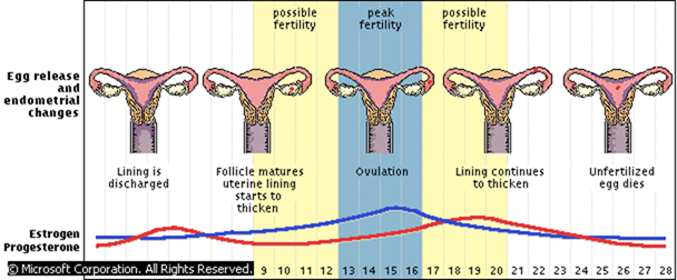
Menstruation
- An average menstrual cycle begins with three to five days of menstruation, the shedding of the uterine lining, during which hormone levels are low.
- At the end of menstruation, pituitary gland secrets FSH which has two functions. It stimulates new Graafian follicles to develop in the ovary and stimulates the ovary to secrete the hormone oestrogen.
- Oestrogen brings about repair and healing of the endometrium, which is destroyed during menstruation.
- Oestrogen accumulates to levels, which stimulate the release of LH. LH stimulates the maturity of Graafian follicle. The mature Graafian follicle releases the ovum into the fallopian tube. This is called Ovulation and occurs on the 14th day.
- The empty Graafian follicle forms the corpus luteum, an endocrine body that secretes progesterone.
- LH stimulates corpus luteum to secrete hormone progesterone. This hormone stimulates thickening and increased blood supply to the endometrium preparing the endometrium for implantation.
- If fertilization takes place, the level of progesterone increases and thus inhibits FSH from stimulating the maturation of another Graafian follicle.
- If fertilization does not take place, the corpus luteum dies and progesterone hormone levels fall.
- Without hormonal support, the uterine lining disintegrates and discharges, beginning a new menstrual period and cycle.
- This cycle lasts for 28 days in human beings.
Assignment
Sanitary Health
- Menopause
- STI
Advantages of Asexual reproduction
- Good qualities from the parents are retained since there is no variation.
- There is faster maturation.
- Its independent of processes such as pollination, fertilisation and fruit and seed dispersal
- New offspring’s are able to obtain nourishment from their parents and are therefore able to survive under unsuitable conditions.
- There is no wastage of a large number of offspring’s.
Disadvantages
- Reduction in strength and vigour in offsprings.
- Undesired qualities are easily inherited.
- Due to faster maturation there are chances of overcrowding and competition.
- Offsprings may not withstand changing environmental conditions due to lack of variation.
Advantages of sexual reproduction
- There is hybrid vigour due to mixing of genetic material.
- There is high adaptability
- Variation form basis for evolutionary changes.
Disadvantages
- May produce individuals with undesirable qualities.
- Method is dependent of union of gametes and therefore may not take place if the two organisms are isolated