Excretion-Process by which living organisms separate and eliminate waste products formed during metabolic processes from the body. They include; carbon IV oxide, excess water and mineral salts, nitrogenous wastes etc. accumulation of these substances may become toxic to cells.
Homeostasis-This is the maintenance of internal environment of cells under constant conditions E.g. temperature, osmotic pressure, blood sugar and chemical constituents.
Egestion. – This is the removal of undigested and indigestible materials from the alimentary Canal of animals.
Secretion. – This is the release of certain useful substances produced by cells e.g. hormones, Enzymes, sebum, saliva and mucus.
Excretion in Plants
- Plants do not have complex organs for excretion because;
- There is very little accumulation of toxic wastes such as nitrogenous wastes.
- Some waste products are re-used in the same plant such as Co2, oxygen and water.
- Some of these gases are removed by simple diffusion through the stomata and lenticels.
- Some plants store wastes in their tissues in non-toxic forms such as nicotine, caffeine, tannins, resins, quinine, morphine etc.
Economic Importance of Plant Excretory Products
- Tannins – They are deposited in dead tissues of wood and barks of trees e.g. in acacia and wattle tree. Tannin is used to treat leather.
- Caffeine – it is stored in coffee berries and tea leaves. It is used as a stimulant.
- Quinine – it is stored in the leaves of aloe and bark of cinchona tree. It is used in the treatment of malaria.
- Cocaine – it is obtained from the leaves of coca plant and is used as an anesthetic.
- Cannabis – found in the leaves and flowers of Cannabis sativa (bhang). It is used to manufacture some drugs.
- Nicotine – found in leaves of tobacco plant and is used in the manufacture of insecticides and narcotic drugs. It also manufactures cigarettes.
- Rubber – it is made from latex of rubber plant. It is used in shoe industry and manufacture of chewing gum.
- Colchicines – it is used in plant breeding and treating of cancer.
- Pappain– it is obtained from raw paw paw and it is used as a meat tenderizer.
- Khat/miraa – it’s chewed and acts as a mild stimulant.
Excretion and Homeostasis in Unicellular Organisms
- Most simple organisms such as the protozoa (amoeba and paramecium) live in aquatic environment.
- They depend mainly on diffusion as the means of excretion.
- Their bodies have a large surface area to volume ratio providing a large surface area for gaseous exchange and excretion to take place by simple diffusion.
- Waste products diffuse from the cytoplasm where they are highly concentrated across the cell membrane into the surrounding water where their concentration is low.
- The organisms also use the contractile vacuole to achieve excretion.
- Amoeba and paramecium live in an aquatic environment that is hypotonic to their body fluids. Water therefore tends to move into their cytoplasm by osmosis.
- The excess water and dissolved chemicals accumulate in the contractile vacuole which releases them to the surrounding water.
Diagram
Excretion in Mammals
- Mammals have an elaborate excretory system since their bodies are complex.
- The main excretory organs in mammals include; lungs, skin, kidneys and the liver.
A Structure and Function of the Mammalian Skin
- Skin is the largest body organ covering the whole body surface.
- It has the following functions.
- Protection of the underlying tissues from entry of micro-organisms, physical damage and ultra violet rays from the sun.
- Regulation of body temperature.
- Excretion of salts, excess water and traces of urea.
- Reception of stimuli such as heat, cold, pain, touch and pressure.
- Synthesis of vitamin D.
- Storage of fats.
Diagram
- The skin is made up of two layers;
- Epidermis (upper and outer layer)
- The dermis (inner layer)
a) Epidermis (upper and outer layer)
- It is made up of three other layers;
- Cornfield layer.
- Granular layer.
- Malphigian layer.
- Cornifield layer
- The Cornifield layer of the epidermis consist of dead cells which form a tough outer coat; that protects the skin against mechanical damage, bacterial infection and water loss;
- Granular layer
- It’s the middle layer of the epidermis and is made up of living cells that give rise to the Cornifield layer.
- Malphigian layer
- Malphigian layer consists of actively dividing cells that contain fine granules of melanin; that prevents the skin against ultraviolet light rays from the sun; melanin gives the skin its colour.
- The Dermis (inner layer)
- It is thicker than the epidermis and consists of the following structures;
- Sebaceous glands produce an oily secretion sebum which give hair its water repelling property; that keeps the epidermis supple and prevents it from dying
Sebum also prevents bacterial attack due to its antiseptic property;
- Has blood vessels; that dilate and contract;
In hot conditions, they dilate; increasing blood flow near the skin surface enhancing blood flow near the skin surface; minimizing heat loss;
Blood vessels supply nutrients and oxygen to skin tissues and also remove waste products and carbon IV oxide.
- Has Hair follicle ;hairs stand during cold weather thus trapping a layer of air which prevents heat loss; In hot weather they lie close to the skin surface; to enhance heat loss to the atmosphere.
- Have many sensory neurons which detects environmental changes; increasing sensitivity of the skins.
- Has subcutaneous layer; contains fat which acts as a heat-insulating layer and a fuel storage.
- Lymphatic vessels; they drain excess tissue fluid.
- Sweat glands; are involved in temperature regulation through loss of excess heat by the evaporating water.
Sweat also excretes excess water, mineral salts, urea and lactic acid.
B The Lungs
- They are involved with the removal of carbon VI oxide which is released by cells during their metabolism.
- Carbon IV oxide would be toxic if it was left to accumulate in the tissues.
C Structure and Function of the Kidney
Diagram fig. 4.3; generalized urinary system of a mammal (page 88 KLB)
- Mammals have a pair of kidneys which are bean shaped and dark red in colour.
- The kidneys are surrounded by a layer of fat which cushions them against mechanical injury.
- Above each kidney are the adrenal glands which secrete hormones.
- Renal artery supplies blood to the kidneys and the renal vein removes the blood.
- Ureter transports urine from the kidney to the bladder which temporarily stores the urine.
- The mammalian kidney has three distinct regions; cortex, medulla and pelvis.
Diagram fig. 4.4(a) and 4.4(b) (page 89 KLB)
Cortex
- It is the outermost region and is dark red in colour.
Medulla
- It is red in colour and extends to form conical structures called pyramids.
- Pyramids open up into the pelvis.
Pelvis
- It’s white in colour and narrows down to form the Ureter.
- The human kidney contains urinary tubules called the nephrons.
Nephron
- It is the basic functional unit of the kidney. Each nephron is made up two main parts;
- Renal tubule
- Glomerulus.
Diagram fig. 4.6. The structure of the kidney nephron
The renal tubule has 5 main parts.
- Bowman’s capsule
- Proximal convoluted tubule
- Loop of Henle
- Distal convoluted tubule
- Collecting tubule
- Bowman’s capsule
- It is a thin walled and cup shaped structure which contains the glomeruli.
- Glomerulus is a fine network of blood capillaries enclosed by the Bowman’s capsule.
- It is made the afferent and efferent arterioles.
- Blood entering the kidney via the renal artery is rich in nitrogenous wastes such as urea.
- Also it has dissolved food substances, plasma proteins, mineral ions, hormones and oxygen.
- Afferent arteriole entering the Glomerulus is wider than the efferent arteriole leaving it.
- This creates extremely high pressure in the Glomerulus coupled with the fact that renal artery branches directly from the aorta where blood is at high pressure.
Diagram: structure of the nephron
- Due to the high pressure in the glomeruli, the liquid part of the blood and dissolved substances of low molecular sizes including urea, glucose, salts and amino acids are forced out of the Glomerulus into the cavity of the Bowman’s capsule.
- The large sized molecules in the plasma such as proteins and blood cells are not filtered out.
- This is because the capillary walls of the Glomerulus and bow mans capsule have very small pores.
- This process is known as ultra-filtration and the filtrate formed is called glomerular filtrate.
- The filtrate then enters the proximal convoluted tubule.
Diagram of ultra-filtration at the Glomerulus
- Proximal convoluted tubule
- As the filtrate flows along the renal tubules, most of the filtered substances in the glomerular filtrate useful to the body are selectively reabsorbed back into the blood.
- The following substances are actively reabsorbed using energy in the proximal convoluted tubule; All glucose, Amino acids and Mineral salts.
- The proximal convoluted tubule is adapted in the following ways for efficient re-absorption of these substances.
- Presence of mitochondria in the cells lining to provide with energy required
- Cells of the tubule have micro-cilli (infoldings) which increase surface area for re-absorption.
- Tubule is long and coiled to increase the surface area.
- Coiling of the tubule reduces the speed of flow of filtrate giving more time for efficient re-absorption.
- Tubule is well supplied with blood capillaries.
- Loop of Henle
- This is where particularly sodium chloride is actively reabsorbed into the blood.
- Loop of Henle has counter current flow between the flow of filtrate and the flow of blood i.e. the filtrate and blood flow in opposite directions.
- The hormone Aldosterone secreted by the adrenal glands regulate the absorption of sodium salts.
- Low content of sodium salts in the blood stimulates adrenal glands to secret more Aldosterone hormone and therefore more salts are reabsorbed from the filtrate.
- Distal convoluted tubule
- When the filtrate reaches here, some water is reabsorbed into the blood by osmosis.
- This is made possible by the following;
– Active intake of sodium salt into the blood at the loop of Henle increases the osmotic potential of the blood.
– The antidiuretic hormone (ADH) secreted by the pituitary gland. ADH increases the permeability of the tubule and blood capillaries to water
- When there is excess water in the body there is less production of ADH and less water is reabsorbed hence production of large amounts of dilute urine.
- If the body has lost a lot of water such as through sweating, this raises the osmotic pressure of blood. Pituitary gland releases more ADH which increases permeability of the kidney tubules to water. More water is reabsorbed hence production of little but concentrated urine.
- The distal convoluted tubule has large surface area, it is has a wall that is one cell thick and is surrounded by may blood capillaries.
- Collecting tubule
- The filtrate in the collecting tubule becomes the urine and moves to the collecting duct.
- Urine flows into the pelvis via the pyramids and is finally emptied into the urinary bladder through the ureter. About 1-2 litres of urine are formed in a day.
- About 250ml of urine in the urinary bladder initiates the urge to urinate. The sphincter muscles relax and urine pass.
Urine Composition
| Substance | % Composition. |
| Water | 95% |
| Urea | 2% |
| Uric acid | 0.03% |
| Creatine | 0.1% |
| Salts | 1.4% |
| Ammonia | 0.04% |
| Proteins | 0% |
| Glucose | 0% |
- The quantity and concentration of the urine in animals is affected by
- Physiological adaptations.
- Habitat of an organism e.g. terrestrial, desert or aquatic.
- Structural adaptations of the animals e.g. a desert rat has long kidney tubules to increase water reabsorption.
Study Questions. Page 93.
Comparison Between Aquatic and Desert Animals
| Fresh Water Animals | Desert Animals. |
| Have many glomeruli to increase ultrafiltration. | Few glomeruli to reduce ultrafiltration. |
| Short loop oh Henle to reduce water reabsorption. | Long kidney tubules to increase water reabsorption. |
| Produce large quantity of dilute urine. | Produce small quantity of concentrated urine. |
Comparison of Composition of urine with that of Glomerular Filtrate and Blood Plasma.
| Substance | % Composition of; | ||
| Plasma | Glomerular Filtrate. | urine | |
| Urea | 0.03 | 0.03 | 2.0 |
| Uric acid | 0.005 | 0.005 | 0.03 |
| Ammonia | 0.001 | 0.001 | 0.004 |
| Glucose | 0.1 | 0.1 | 0 |
| Amino acids | 0.05 | 0.05 | 0 |
| Mineral salts | 0.70 | 0.70 | 1.4 |
| Blood proteins. | 8.00 | 0 | 0 |
Functions of the kidney include:
- Excretion.
- Osmoregulation.
- Ionic balance.
- Regulation of PH
Kidney Diseases
- Nephritis
This is the inflammation of the glomerulus of the kidney. It is caused by bacteria or infections such as small pox and measles.
Symptoms
- Headaches and vomiting
- Fever
- Passing coloured urine
- Presence of proteins in urine
Treatment
- Use of antibiotics
- Use of just adequate amounts of salts and proteins in diets Kidney stones
Causes
- Lack of vitamins such as vitamin A and inadequate intake of water
- Chemical salts in urine that crystallize to form hard stones.
Symptoms
- Increased frequency in passing out urine
- Pain and soreness in the upper backside
- Difficulty in passing out urine
- Fever
Control & Treatment
- Seeking medical assistance
- Taking a balanced diet with adequate amount of water and vitamins
- Dialysis or artificial washing out of the wastes
- Use of laser beam to disintegrate the stones
- Kidney transplant
- Kidney failure
- This is the failure of the kidney to perform as a result of a drop in blood pressure due to heart failure, haemorrhage or shock.
- If failure is due to other causes, the condition can be corrected by;
- Kidney dialysis
- Kidney transplant
- Albiminuria (Proteins in Urine).
- This is also called Proteinuria
- Capillaries of the glomerulus lose their ability to be selective and start allowing blood proteins to pass through into the kidney tubules. These proteins are released in urine.
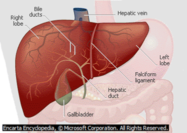
D The Liver and its Structure
- This is the second largest excretory organ after the skin. It receives blood from two blood vessels; the hepatic portal vein from the alimentary canal and hepatic artery from the aorta.
Homeostatic Functions of the Liver
Regulation of blood sugar level
- Excess glucose is converted to glycogen ;and stored in the liver under the influence of the hormone insulin secreted by the pancreas. Another hormone called glucagon; stimulates the conversion of glycogen to glucose; when there is shortage of glucose in the body; Glucagon is also secreted by the pancreas
- Deamination
- The liver breaks down excess amino acids; The amino group is removed as ammonia which is toxic;
- Ammonia is changed into urea which is less toxic in the ornithine cycle.
| Ornithine Cycle Enzyme arginase |
2NH3 + CO2 CO(NH2)2 + H20
Ammonia Carbon iv Urea Water
(Toxic) Oxide (less toxic)
- The remaining carbon skeleton oxidized to carbon IV oxide and water; this process leads to release of energy. The carbon skeleton may be converted to glucose to be used during respiration;
- Detoxification
- Toxic substances are made harmless in the liver e.g.
- Ammonia from the process of deamination is converted in the liver into urea; which is less toxic.
- Bacterial toxins are converted to less toxic substances by liver cells;
- Hydrogen peroxide produced by respiring cells is broken down into water and oxygen which are harmless by the enzyme catalase found in the liver.
| Enzyme Catalase |
Hydrogen Peroxide Water + Oxygen
(H2O2) (H2O) (O2)
- Regulation of plasma proteins
- The liver produces most of the proteins found in blood; fibrinogen and prothrombin which play a role in blood clotting. Albumin and globulins are also produced by the liver. Globulins act as antibodies;. Albumin contributes to the maintenance of osmotic pressure in the body; Non essential amino acids are synthesized by the liver;
- Storage of vitamins A, B,D,E and K and mineral salts
- The liver store vitamins A, B, D, E and K. Iron released from the breakdownof erythrocytes is stored in the liver cells; in the form of a compound called ferritin. The liver therefore is a good source of these vitamins and iron;
- Heat production (Thermoregulation)
- The various metabolic activities of the liver lead to release of heat energy; This energy is distributed by the blood to other parts of the body hence contributing to maintenance of constant body temperature;
- Inactivation of hormones and drugs
- After performing their functions, hormones and drugs are chemically modified to inactive compounds; The by-products are eliminated through the kidneys and faeces and via bile;
- Storage of blood
- The large size and high capacity for contraction and expansion of its veins enables the liver to hold a large volume of blood; It therefore regulates the volume of blood in the general circulation depending on the body’s requirements ;
- Regulation of cholesterol and fat metabolism
- When carbohydrates are in short supply in the body, fats in different parts of the body are mobilized and taken to the liver; The fats are oxidized to carbon (IV) oxide and water with the production of energy or modified and sent to tissues for oxidation;
- Manufacture of red blood cells in foetus.
Liver Diseases and Disorders
- Liver Cirrhosis
- This is the hardening of the liver tissues due to death of liver cells.
- This is caused by ingestion of toxic chemicals such as alcohol.
- Bacteria, viruses and parasites such as liver flukes can also cause the disease.
Control
- Avoid excess alcohol.
- Avoid fatty diets.
- Low salt intake
- Hepatitis
- This is a viral disease causing inflammation of the liver.
- It is transmitted through contaminated food, milk and water.
- There are two types of hepatitis; Hepatitis A and B.
- Jaundice
- This is characterized by the yellowing of the skin due to the failure of the liver to excrete bile.
Homeostasis
- This is the maintenance of internal environment of cells under constant Conditions E.g. temperature, osmotic pressure, blood sugar and chemical constituents.
Principles of Homeostasis
- Various body systems such as circulatory, excretory, endocrine (hormonal) and nervous work in a coordinated way to bring about homeostasis.
- These systems work on a feedback mechanisms. There are two types of feedback mechanisms.
Negative Feedback Mechanism
When a factor in the body such as temperature or blood sugar level falls below normal or rises above the normal, it is detected and corrected via the negative feedback mechanism.
Such an action is through:
An increase in the level if it is dropping
A decrease in the level if it is increasing
This restores the condition to the normal.
Positive Feedback Mechanism
In positive feedback mechanism, a change below or above the normal is not corrected.
The following are some of the factors regulated through homeostasis.
- Temperature
- Osmoregulation (water and salt balance)
- Ionic content regulation
- Blood sugar regulation
- Temperature Regulation. (Thermoregulation)
- Hypothalamus of the brain is the thermoregulatory center. It also controls other homeostatic processes such as Osmoregulation, and blood sugar level.
Skin and Thermoregulation
The skin is adapted in the following ways to effect thermoregulation
- It has Hair shaft;
Connected to erector pili muscles;
In low Temperature Erector pili muscle contract raising hair shaft erect;
Hair traps air which insulates the body/poor conductor of heat.;
In high temperature, the Erector pili muscle relax and extends;
Hair shaft lies on the skin;
Little or no air is trapped;
Skin loses heat through convection /conduction /radiation ;
2. Blood vessels
In High temperature they vasodilate;
More blood flows near skin surface;
Heat is lost through conduction /convection/ radiation;
In Low temperature they Vasoconstrict;
Little blood flows near the skin;
Less heat or ho heat lost through conduction/convection/ radiation;
Diagrams
3) Sweat gland
- In High temperature, Sweating occurs and ( evaporates)andCarries latent heat of vaporization; cooling the body;
4) Has subcutaneous layer;
contains fat which acts as a heat-insulating layer. Organisms in cold areas have thick subcutaneous layer for heat insulation.
Homoiotherms and Poikilotherms
Homoiotherms (Endotherms)
- They are the animals whose body temperature is maintained at a constant body temperature despite the wide fluctuations in the temperature of the external environment e.g. birds and mammals.
Poikilotherms (Ectotherms)
- These are organisms with variable body temperature according to the temperature of the local atmosphere e.g. in organisms such as reptiles, amphibians, insects, and fish.
Methods of Regulating Body Temperature in Animals.
- Metabolic activities of the Body, such as shivering to raise body temperature.
- Insulatory mechanisms such as dilation or constriction of blood vessels, hair movement etc.
- Behavioral activities such as clustering together, burrowing, basking, hibernation, aestivation, putting on warm clothes etc.
- Presence of adaptive features such as hair/fur, subcutaneous tissue etc.
Hibernation is where animals go into deep sleep for long period of time due to cold.
Aestivation is where animals go into deep sleep due to dry and harsh conditions.
Differences Between Homoiotherms and Poikilotherms.
| Poikilotherms | Homoiotherms |
| They are sluggish under cold conditions. | They remain active even under cold conditions. |
| They hibernate to avoid death by freezing under very cold conditions. | Only the small animals hibernate because they have large surface area to volume ratio hence lose a lot of heat. |
| They aestivate under very hot conditions. | They do not aestivate because they can maintain constant body temperature. |
| They are easy prey to predators due to their hibernation and aestivation. | Not easy to prey because they active always. |
| Require less food because they get heat from the environment to warm their bodies. | Require more food because they use it to generate heat for maintaining the temperature constant. |
- Osmoregulation (Water and Salt Balance).
- The osmotic pressure of the body fluids must be kept at a constant so as to have a favourable environment for the normal functioning of cells. This is determined by the relative amounts of water and solutes (salts) in the body fluids.
- If the osmotic pressure of these fluids falls below that of the cells, the cells take in water by osmosis, swell and may burst.
- If the osmotic pressure of thee fluids was higher than that of the cells, the cells would lose water and shrink.
- The hypothalamus and the Pituitary gland are involved in Osmoregulation in the following ways;
- When the osmotic pressure of the blood rises due to dehydration, the hypothalamus is stimulated and sends an impulse to the pituitary gland which secretes the Antidiuretic Hormone (ADH) or Vasopressin into the blood. ADH increases permeability of the kidney tubules to water. More water is reabsorbed, osmotic pressure of the blood falls hence production of little but concentrated urine.
- When osmotic pressure of the blood falls due to excess water in the body there is less production of ADH and less water is reabsorbed hence production of large amounts of dilute urine.
Diabetes Insipidus
- This is a condition whereby large quantities of dilute urine are produced when the pituitary gland fails to produce ADH or produces it in inadequate amounts. This condition is also known as Diuresis.
- Regulation of Ionic Content
- Important ions must be regulated within narrow ranges for efficient functioning of the cells.
- Ions are involved in processes such as respiration, protein synthesis, muscle contraction etc.
- The level of sodium ions is regulated by a hormone called Aldosterone produced by the adrenal glands.
- When the level of sodium ions is low in the blood, more Aldosterone is released which stimulates reabsorption of sodium ions into the blood.
- If sodium ions concentration in the blood rises above the optimum level, adrenal glands produce less Aldosterone into the blood and less amounts of sodium ions are reabsorbed.
- Regulation of Blood Sugar Level.
- All sugars such as galactose, lactose and fructose are converted to glucose.
- Glucose is broken down to release energy and excess is converted into glycogen and stored in the liver or converted into fats as stored as adipose tissue.
- Some glucose flows in general circulation of blood and is maintained within a narrow range of 90-100mg per 100cm3 of blood.
- The pancreas produces two hormones Insulin and Glucagon that are responsible for blood sugar regulation.
- When there is excess sugar in the blood, insulin is produced and regulates the blood sugar level by the following;
- Converts excess glucose into glycogen for storage.
- Inhibits conversion of glycogen to glucose.
- Converts glucose into fats.
- Increases breakdown of glucose to release energy.
- When the level of the blood sugar falls, glucagon is secreted and corrects the situation by the following;
- Increases the breakdown of glycogen into glucose.
- Increases the conversion of fats and proteins into glucose.
- Inhibits the conversion of glucose into energy.
NB/. The hormone adrenaline produced by the adrenal glands also has homeostatic effect on glucose.
It is released during emergencies to avail glucose for fight or flight.
Diabetes Mellitus (Sugar Disease)
- This is due to a deficiency in insulin secretion from the pancreas.
- This leads to very high levels of sugar in the blood that cannot be utilised by cells hence eliminated by kidney in urine.
Symptoms
- Presence of glucose in urine
- Loss of body weight due to breakdown of fats and proteins
- Chronic starvation
- Thirst sensation.
Control
- Insulin injection into the blood stream
- Avoid foods rich in sugars.
- Avoid excessive intake of alcohol.
Question
- Explain why insulin is not administered orally (through the mouth)